
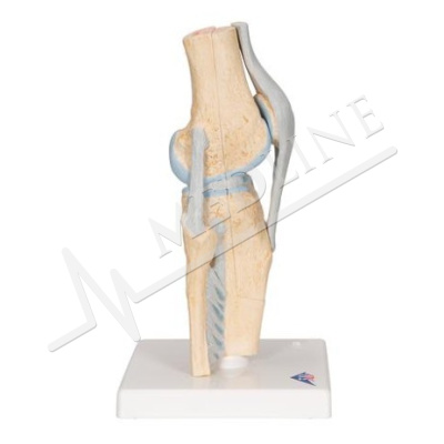
Foot skeleton model with ligaments and muscles M 34/1
Description
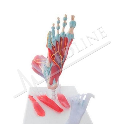
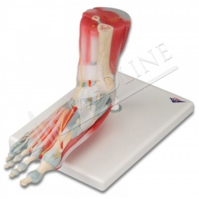
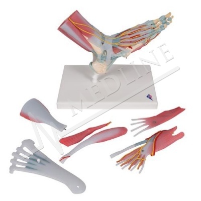
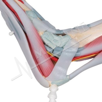
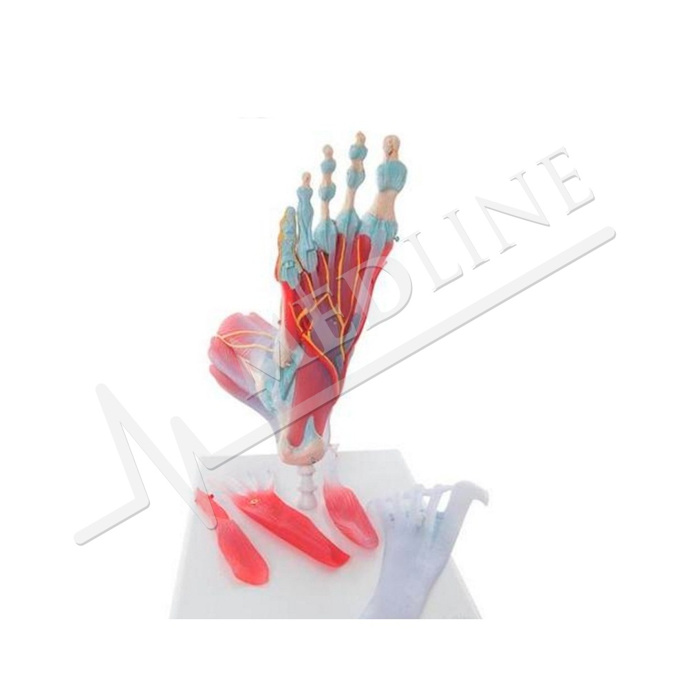
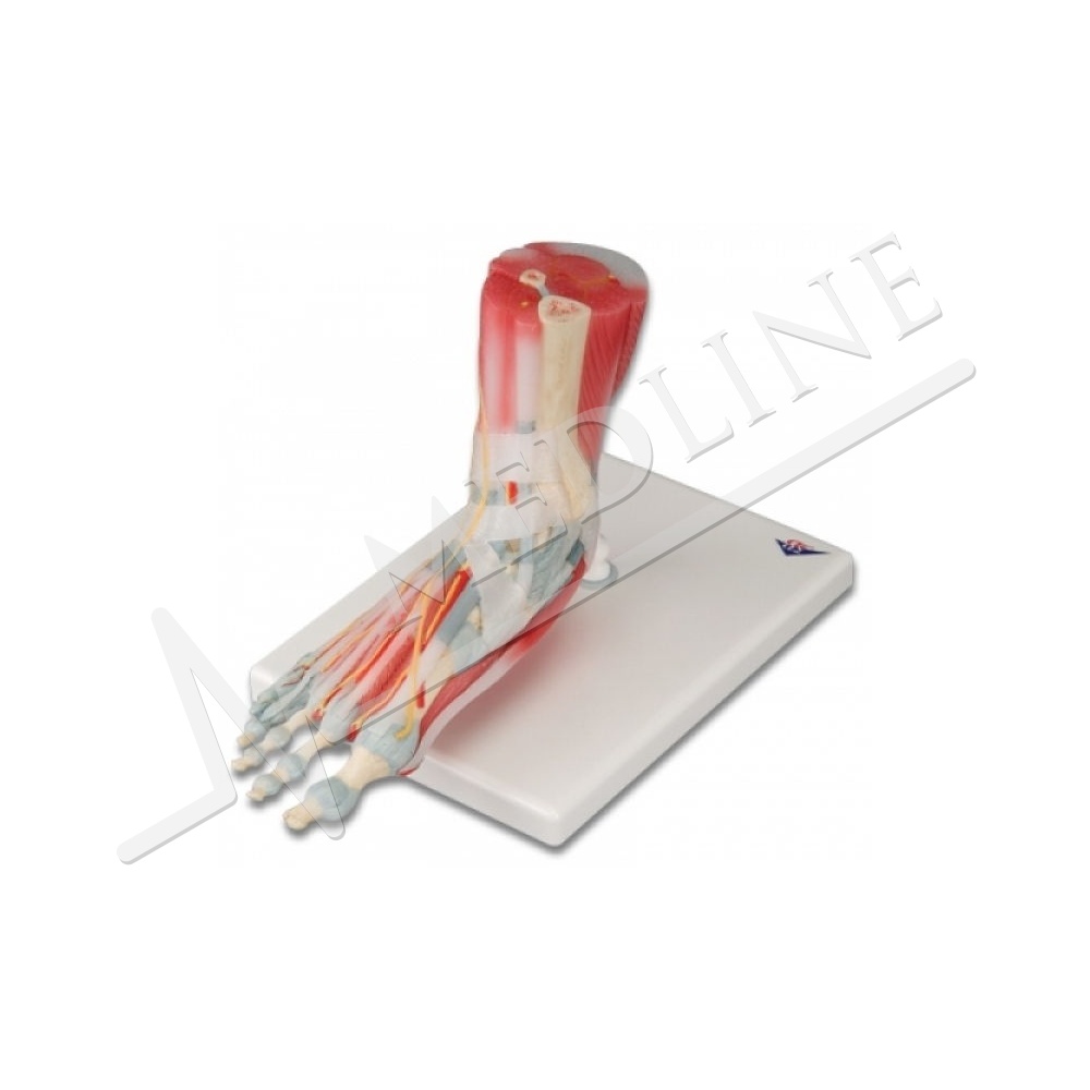
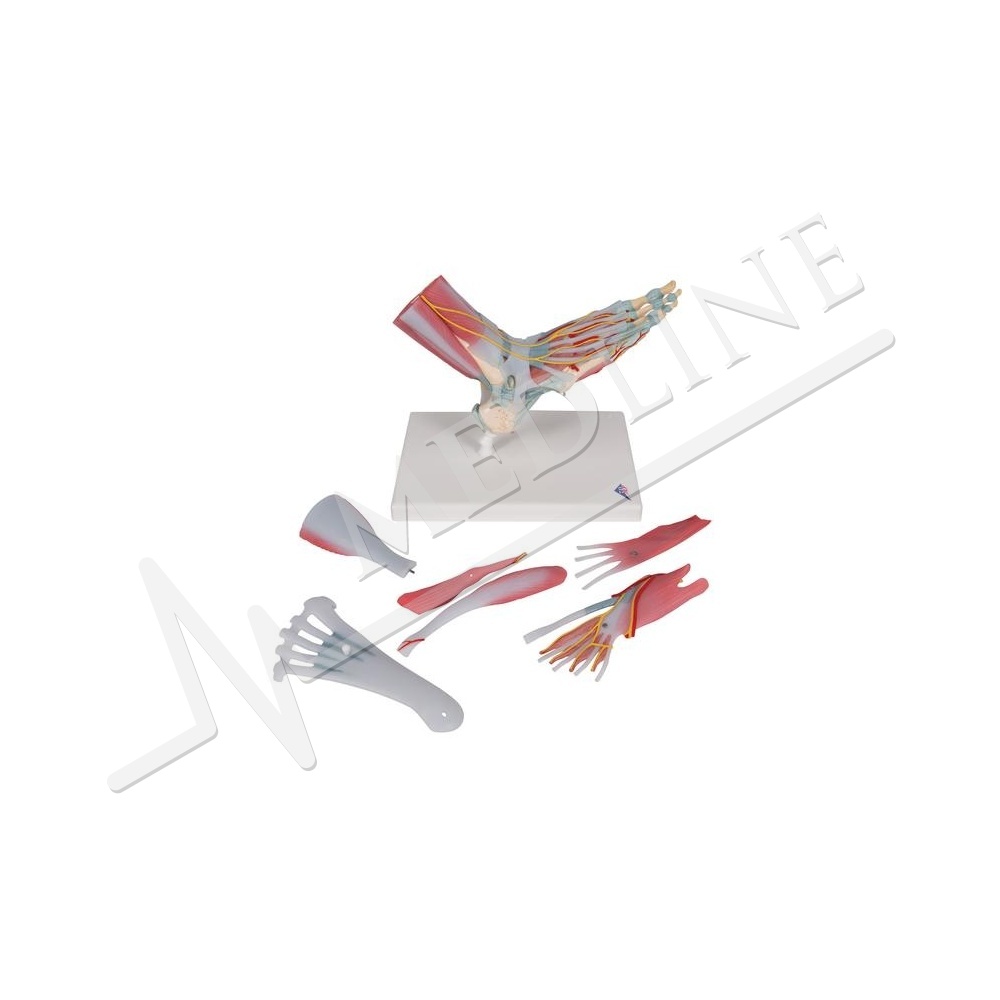
This anatomically detailed model of the foot and lower leg can be disassembled into six removable pieces for a detailed study of the area. This model presents not only bones but also muscles, tendons, ligaments, nerves, arteries and veins.
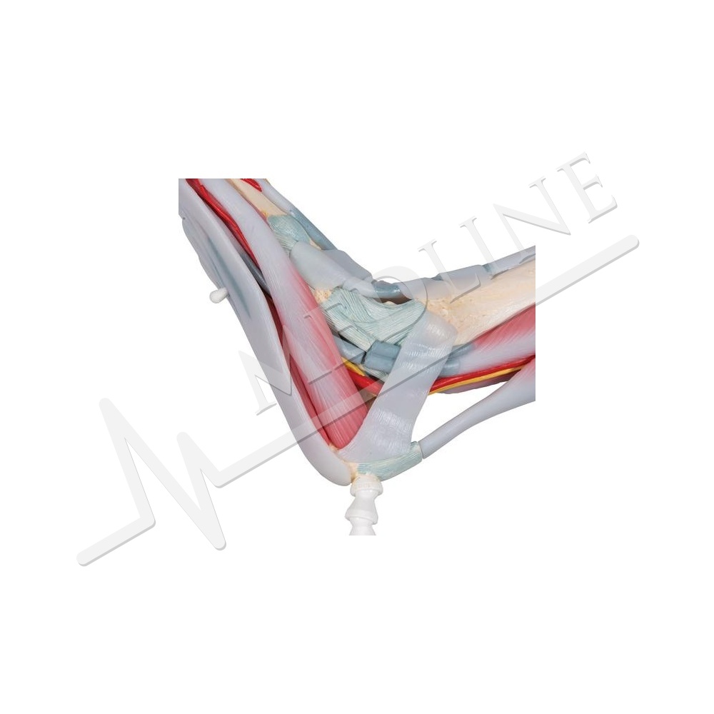
The frontal view shows the extensor muscles of the lower leg. The tendons can be followed as they pass under crucial transverse and cruciate ligaments, all the way to the insertion points. In addition, all tendon sheaths are visible.
On the dorsal part of the model, the gastrocnemius muscle can be removed to reveal the deeper anatomical elements. The sole of the foot is represented in three layers, the first presenting the flexor tendons
on point. This muscle can be removed to reveal the sole of the foot, the tendon of the long
flexor of the finger and the flexor hallucis muscle. This second layer can then be removed to reveal deeper anatomical details.
This model is very complete in terms of quality and value.
Every original 3B Scientific® Anatomy Model gives you direct access to its digital twin on your smartphone, tablet or desktop device.
Enjoy using the exclusive virtual anatomy content with the following features:
Freely rotate your digital model and zoom in and out
Display hotspots and their anatomical structures
Augmented Reality (AR) feature starts your virtual anatomy model
Anatomy Quiz function to test and improve your anatomical knowledge with instant results and final score evaluation
Drawing function that allows image customization with save and share function
Useful Notes function to help you with your personal learning
Possibility to learn both male and female anatomy
Easy access to 3D content both online and offline
Available in 11 languages
To get started, simply scan the QR-code located on your 3B Scientific® Anatomical Model, download the new 3B Smart Anatomy app and step into the virtual world of Human Anatomy
Mass: 1.2 kg
Dimensions: 23x26x19