

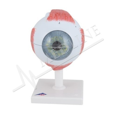
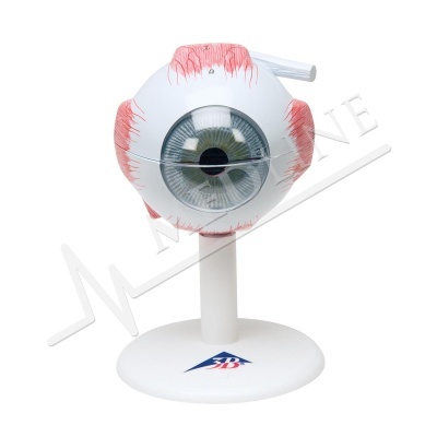
Microanatomy Eye F16
Description
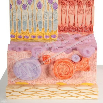
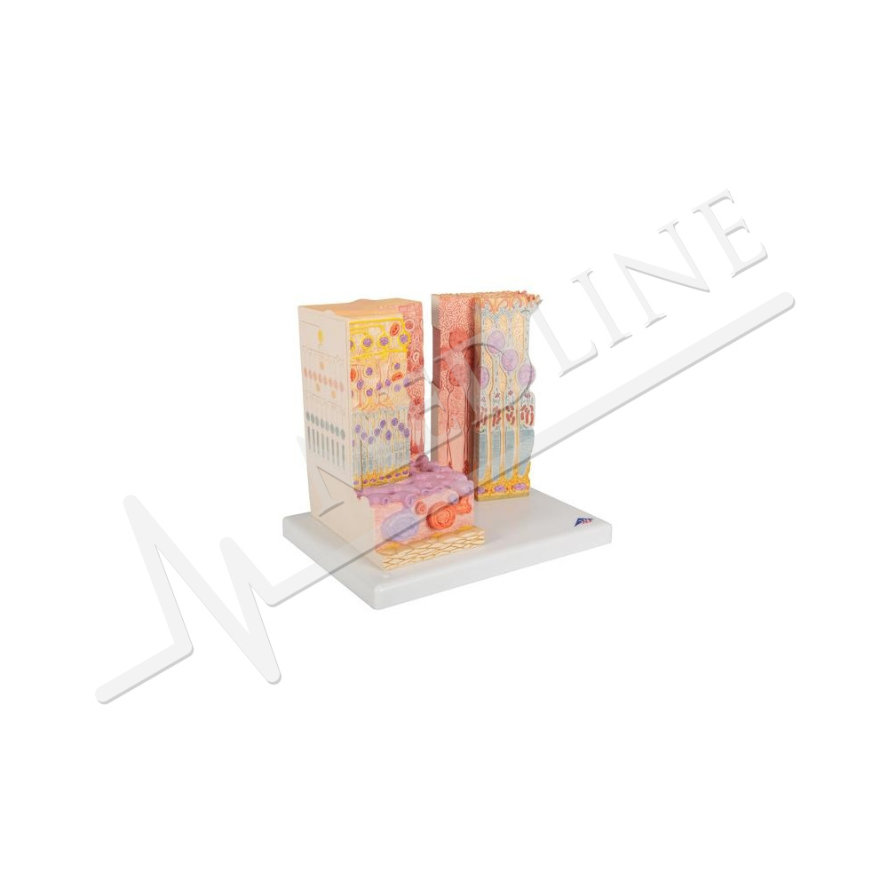
The MICROanatomy™ Eye model illustrates the microscopic anatomical structure of the retina with choroid and sclera. The left block-like, layered side of the eye model shows the complete structure of the retina including the supplying vascular layer and parts of the sclera from a light microscopic view.
The right part of the eye model is a sectional enlargement. MICROanatomy™ Eye shows the microscopic structure of the photoreceptors and the cells of the pigmented layer.
Left part of MICROanatomy™ Eye 850-times enlarged - right part 3800-times enlarged. You've never seen the human eye like this before!
Every original 3B Scientific® Anatomy Model gives you direct access to its digital twin on your smartphone, tablet or desktop device.
Enjoy using the exclusive virtual anatomy content with the following features:
Freely rotate your digital model and zoom in and out
Display hotspots and their anatomical structures
Augmented Reality (AR) feature starts your virtual anatomy model
Anatomy Quiz function to test and improve your anatomical knowledge with instant results and final score evaluation
Drawing function that allows image customization with save and share function
Useful Notes function to help you with your personal learning
Possibility to learn both male and female anatomy
Easy access to 3D content both online and offline
Available in 11 languages
To get started, simply scan the QR-code located on your 3B Scientific® Anatomical Model, download the new 3B Smart Anatomy app and step into the virtual world of Human Anatomy.
Weight 1.11 kg
Dimensions 25 x 23 x 18.5 cm